
Left Leg & Popliteal Fossa
Purpose
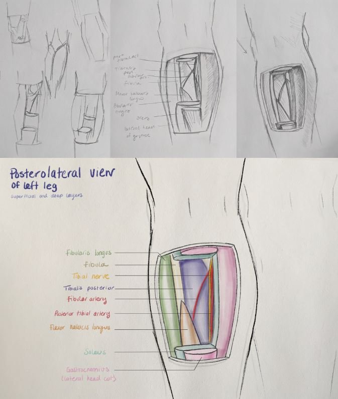
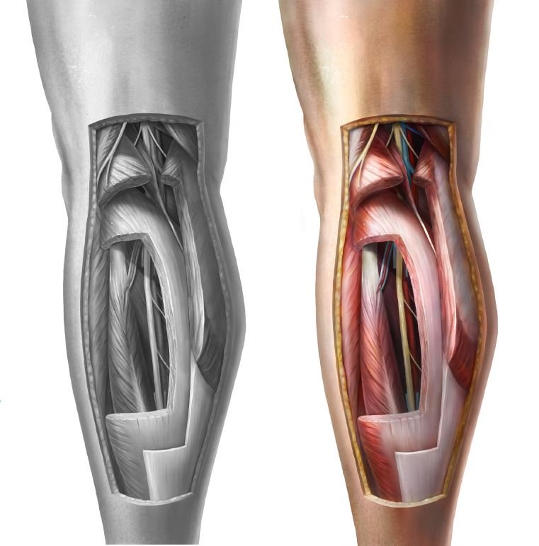
To show the relationship between the superficial and deep layers of the posterior compartment of the left leg from a non-traditional viewpoint.
Clients
Dave Mazerski, Shelley Wall, & Dr. Anne Agur
Date
April 2021
Medium
Textbook Illustration
Tools
Photoshop, Procreate, Illustrator, 3D Slicer, Zbrush
Primary Audience
Anatomy students
Ideation
The project began with quick and rough sketches of potential ideas, which later led to focusing on a piece related to the lower limb. The idea of showing the relationship between the superficial and deep layers of the posterior compartment of the leg interested me as both layers are typically not shown in relation to each other in anatomical illustrations. Additionally, anatomical atlases typically show these layers from a posterior or a posteromedial view, prompting me to explore illustrating these structures from a posterolateral view.

Reference Gathering
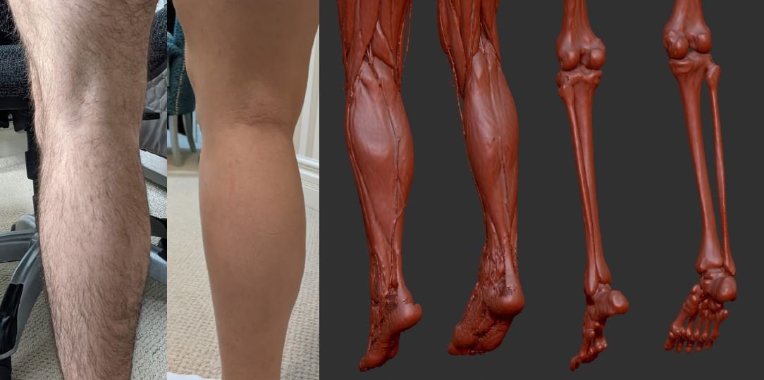
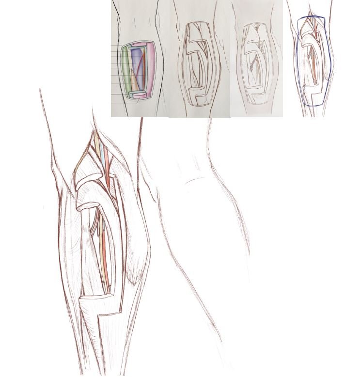
After an initial round of feedback, I chose to extend the cut region up into the popliteal fossa. From this point, I began to gather a variety of references, including images of my family members’ legs in my chosen viewpoint. To help visualize the location of bony landmarks and their relation to the superficial muscles of the leg, I extracted a 3D model from CT data (Melanix DICOM dataset extracted in 3D Slicer and viewed in ZBrush). Illustrations from a variety of anatomical atlases were used to piece together the anatomy of the leg and popliteal region. This included illustrations of the layers of the posterior compartment and lateral compartment, along with cross-sectional illustrations down the leg. These, in combination with the 3D models and leg images, were used to construct the anatomy of the calf region with cuts in gastrocnemius and soleus to reveal the underlying structures. The majority of these references had to be flipped horizontally while being referred to due to them depicting the anatomy of the right leg rather than the left.
Sketch Refinement
After analyzing and studying my references, I went through several rounds of sketches. I consulted with my content expert, Dr. Anne Agur, to review the anatomical accuracy of my illustration. It was also recommended to me that I elimate the right leg from the illustration to put emphasis on the anatomy of the left leg. Instead of including the right leg in the background, I decided that I would include a small line drawing of both legs with the highlighted cut region in my final drawing to help orient the viewer.

Building a Maquette
To understand how light would interact with the cut structures and overall form, I created a maquette of the lower limb using clay. This model was used to test different lighting options. In the end, I landed on using upper left hand light for my maquette and viewed it both with just the cut muscles alone, and with an additional sheath of clay to represent the cut skin.
Gray Scale Base
Once at the stage of creating the final drawing, I began by blocking out all of the structures with flat shapes in grayscale using procreate. These shapes were later used as a base to render on top of using clipping masks.

Texture Reference Gathering
To render the textures of muscles and fat, I watched surgical videos and viewed images of adipose and raw meat. I also used cadaveric images and anatomical illustrations to view how the muscle fibers were oriented, or any other specific features such as the large aponeurosis on the outer surface of soleus. These references were also used in the colourizing stage to mimic the colour variation of these tissues.


Grayscale Rendering
Using both the texture references and my maquette for lighting information, I rendered the illustration in grayscale using Procreate. When rendering, I began with a first overall lighting pass using a soft airbrush. I later experimented with a variety of textured brushes including many of the charcoal, painting, noise, rough skin, and fine hair brushes. I finished the rendering stage by accentuating details with either a gouache or 6B pencil brush and adding highlights with the studio pen.
Colourizing
The grayscale image was later brought into Photoshop to colourize each layer. The previous images used to provide information for texture were also used to provide colour information for all of the tissues. Additionally, images of the legs and thigh that I captured at the beginning of the project were used to add colour to the skin.
References
Agur, A. M., & Dalley, A. F. (2017). Grant's atlas of anatomy (14th ed.). Philadelphia, PA: Wolters Kluwer.
Agur, A. M., & Dalley, A. F. (2019). Moore's essential clinical anatomy (6th ed.). Philadelphia, PA: Wolters Kluwer.
Anson, B.J. (1963). An atlas of human anatomy. Philadelphia, PA: W. B. Saunders Co.
Guangwen, G., & Xu, W. (1991). Colour atlas of human anatomy (1st ed.). Beijing: People's Medical Publishing House.
JBJSmedia. (2017, July 20). Single and dual incision fasciotomy of the lower leg [Video file]. YouTube.
Retrieved April 12, 2021, from https://www.youtube.com/watch?v=WNyKp1hlZKc
Pernkopf, E. (1989). Atlas of topographic and applied human anatomy [Atlas der topographischenund angewandten Anatomie des Menchen]. (H. Monsen, Trans., W. Platzer, Ed.) (3rd ed.). Baltimore, MD: Urban & Schwarzenberg.
Rohen, J. W., Yokochi, C., & Lütjen-Drecoll, E. (2011). Color atlas of anatomy: A photographic study of the human body (7th ed.). Philadelphia, PA: Lippincott Williams & Wilkins.
Schuenke, M., Schulte, E., & Schumacher, U. (2016). Atlas of anatomy (3rd ed.) (A. M. Gilroy & B. R. Macpherson, Eds.). New York, NY: Thieme.
Sinelnikov, R. D. (1963). Atlas of human anatomy in three volumes. Moscow: MIR.
Spalteholz, W. (1967). Atlas of human anatomy. (A. Nederveen & G. N. C. Crawford, Trans., R. Spanner, Ed.) (16th ed.). London: Butterworths.
WUSTL Learn Surgery. (2018, October 1). Tibial nerve decompression at the soleus - extended (feat. Dr. Mackinnon [Video file]. YouTube. Retrieved April 12, 2021, from https://www.youtube.com/watch?v=6AhkmsuKGns